
Functional label-free assessment of fibroblast differentiation in 3D collagen-I-matrices using particle image velocimetry
 a
and
Tilo
Pompe
*a
a
and
Tilo
Pompe
*a
Abstract
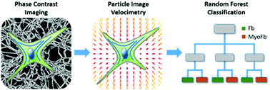
Fibroblasts are a diverse population of connective tissue cells that are a key component in physiological wound healing. Myofibroblasts are differentiated fibroblasts occurring in various physiological and pathological conditions, like in the healing of wounds or in the tumour microenvironment. They exhibit important functions compared to fibroblasts in terms of proliferation, protein secretion, and contractility. The gold standard to distinguish myofibroblasts is alpha-smooth muscle actin (αSMA) expression and its incorporation in stress fibres, which is only revealed by gene expression analysis and immunostaining. Here, we introduce an approach to functionally determine the myofibroblast status of live fibroblasts directly in in vitro cell culture by analysing their ability to contract the extracellular matrix around them without the need for labelling. It is based on particle image velocimetry algorithms applied to dynamic deformations of the extracellular matrix network structure imaged by phase contrast microscopy. Advanced image analysis allows us to distinguish between various differentiation stages of fibroblasts including the dynamic change over several days. We further apply machine learning classification to automatically evaluate different cell culture conditions. With this new method, we provide a versatile tool to functionally evaluate the dynamic process of fibroblast differentiation. It can be applied for in vitro screening studies in biomimetic 3D cell cultures with options to extend it to other cell systems with contractile phenotypes.
 Please wait while we load your content...

