
A 3D pancreatic tumor model to study T cell infiltration†
 a
Yi Juan
Teo,
b
Alrina Shin Min
Tan,
b
Damien Zhi Ming
Tan,
c
Paolo
Decuzzi,
a
Andrea
Pavesi
c
and
Giulia
Adriani
*bd
a
Yi Juan
Teo,
b
Alrina Shin Min
Tan,
b
Damien Zhi Ming
Tan,
c
Paolo
Decuzzi,
a
Andrea
Pavesi
c
and
Giulia
Adriani
*bd
Abstract
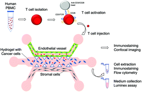
The desmoplastic nature of the pancreatic ductal adenocarcinoma (PDAC) tumor microenvironment (TME) prevents the infiltration of T cells and the penetration of chemotherapeutic drugs, posing a challenge to the validation of targeted therapies, including T cell immunotherapies. We present an in vitro 3D PDAC-TME model to observe and quantify T cell infiltration across the vasculature. In a three-channel microfluidic device, PDAC cells are cultured in a collagen matrix in the central channel surrounded, on one side, by endothelial cells (ECs) to mimic a blood vessel and, on the opposite side, by pancreatic stellate cells (PSCs) to simulate exocrine pancreas. The migration of T cells toward the tumor is quantified based on their activation state and TME composition. The presence of EC-lining drastically reduces T cell infiltration, confirming the essential role of the vasculature in controlling T cell trafficking. We show that activated T cells migrate ∼50% more than the not-activated ones toward the cancer cells. Correspondingly, in the absence of cancer cells, both activated and not-activated T cells present similar migration toward the PSCs. The proposed approach could help researchers in testing and optimizing immunotherapies for pancreatic cancer.
 Please wait while we load your content...

