A turn-on fluorescent probe for Lu3+ recognition and bio-imaging in live cells and zebrafish†
Abstract
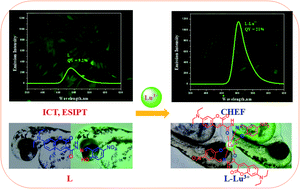
A new Lu3+ selective fluorescent probe L was synthesized and characterized. The optical properties of L were investigated by using absorption and fluorescence spectral studies in 7 : 3 (v/v) aqueous dimethyl sulphoxide. Upon addition of Lu3+ in a pH 4 (acetate buffer) solution of L, the weakly fluorescent probe L became highly fluorescent. The fluorescence intensity increased five-fold at 490 nm with excitation at 437 nm. The formation of 2 : 1 complexation between L and Lu3+ was confirmed by Job's plot. The binding constant (Ka, 1.43 × 10⁴ M−1) was determined by the Benesi–Hildebrand (BH) method. The limit of detection (LOD) of Lu3+ using L was found to be 23 nM. The binding mechanism of L with Lu3+ was studied by 1H-NMR, ESI mass spectroscopy, and theoretical studies. Further, the probe L was successfully used to bioimage Lu3+ in a zebrafish gill cell line (DrG) and in the yolk, papillae of the eyes, and head of zebrafish embryos (Danio rerio), therefore providing a powerful live imaging approach for investigating chemical signaling in complex multicellular systems.


 Please wait while we load your content...