Atomic force microscopy and surface plasmon resonance for real-time single-cell monitoring of bacteriophage-mediated lysis of bacteria†
Abstract
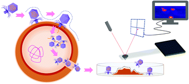
The growing incidence of multidrug-resistant bacterial strains presents a major challenge in modern medicine. Antibiotic resistance is often exhibited by Staphylococcus aureus, which causes severe infections in human and animal hosts and leads to significant economic losses. Antimicrobial agents with enzymatic activity (enzybiotics) and phage therapy represent promising and effective alternatives to classic antibiotics. However, new tools are needed to study phage–bacteria interactions and bacterial lysis with high resolution and in real-time. Here, we introduce a method for studying the lysis of S. aureus at the single-cell level in real-time using atomic force microscopy (AFM) in liquid. We demonstrate the ability of the method to monitor the effect of the enzyme lysostaphin on S. aureus and the lytic action of the Podoviridae phage P68. AFM allowed the topographic and biomechanical properties of individual bacterial cells to be monitored at high resolution over the course of their lysis, under near-physiological conditions. Changes in the stiffness of S. aureus cells during lysis were studied by analyzing force–distance curves to determine Young's modulus. This allowed observing a progressive decline in cellular stiffness corresponding to the disintegration of the cell envelope. The AFM experiments were complemented by surface plasmon resonance (SPR) experiments that provided information on the kinetics of phage-bacterium binding and the subsequent lytic processes. This approach forms the foundation of an innovative framework for studying the lysis of individual bacteria that may facilitate the further development of phage therapy.


 Please wait while we load your content...