
Rapid collagen-directed mineralization of calcium fluoride nanocrystals with periodically patterned nanostructures†

Abstract
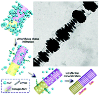
Collagen fibrils present periodic structures, which provide space for intrafibrillar growth of oriented hydroxyapatite nanocrystals in bone and contribute to the good mechanical properties of bone. However, there are not many reports focused on bioprocess-inspired synthesis of non-native inorganic materials inside collagen fibrils and detailed forming processes of crystals inside collagen fibrils remain poorly understood. Herein, the rapid intrafibrillar mineralization of calcium fluoride nanocrystals with a periodically patterned nanostructure is demonstrated. The negatively charged calcium fluoride precursor phase infiltrates collagen fibrils through the gap zones creating an intricate periodic mineralization pattern. Later, the nanocrystals initially filling the gap zones only expand gradually into the remaining space within the collagen fibrils. Mineralized tendons with organized calcium fluoride nanocrystals acquire mechanical properties (indentation elastic modulus ∼25.1 GPa and hardness ∼1.5 GPa) comparable or even superior to those of native human dentin and lamellar bone. Understanding the mineral growth processes in collagen may facilitate the development of tissue engineering and repairing.
 Please wait while we load your content...

