Development of a human neuromuscular tissue-on-a-chip model on a 24-well-plate-format compartmentalized microfluidic device†
Abstract
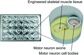
Engineered three-dimensional models of neuromuscular tissues are promising for use in mimicking their disorder states in vitro. Although several models have been developed, it is still challenging to mimic the physically separated structures of motor neurons (MNs) and skeletal muscle (SkM) fibers in the motor units in vivo. In this study, we aimed to develop microdevices for precisely compartmentalized coculturing of MNs and engineered SkM tissues. The developed microdevices, which fit a well of 24 well plates, had a chamber for MNs and chamber for SkM tissues. The two chambers were connected by microtunnels for axons, permissive to axons but not to cell bodies. Human iPSC (hiPSC)-derived MN spheroids in one chamber elongated their axons into microtunnels, which reached the tissue-engineered human SkM in the SkM chamber, and formed functional neuromuscular junctions with the muscle fibers. The cocultured SkM tissues with MNs on the device contracted spontaneously in response to spontaneous firing of MNs. The addition of a neurotransmitter, glutamate, into the MN chamber induced contraction of the cocultured SkM tissues. Selective addition of tetrodotoxin or vecuronium bromide into either chamber induced SkM tissue relaxation, which could be explained by the inhibitory mechanisms. We also demonstrated the application of chemical or mechanical stimuli to the middle of the axons of cocultured tissues on the device. Thus, compartmentalized neuromuscular tissue models fabricated on the device could be used for phenotypic screening to evaluate the cellular type specific efficacy of drug candidates and would be a useful tool in fundamental research and drug development for neuromuscular disorders.


 Please wait while we load your content...