Cross-sectional focusing of red blood cells in a constricted microfluidic channel†

Abstract
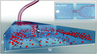
Constrictions in blood vessels and microfluidic devices can dramatically change the spatial distribution of passing cells or particles and are commonly used in biomedical cell sorting applications. However, the three-dimensional nature of cell focusing in the channel cross-section remains poorly investigated. Here, we explore the cross-sectional distribution of living and rigid red blood cells passing a constricted microfluidic channel by tracking individual cells in multiple layers across the channel depth and across the channel width. While cells are homogeneously distributed in the channel cross-section pre-contraction, we observe a strong geometry-induced focusing towards the four channel faces post-contraction. The magnitude of this cross-sectional focusing effect increases with increasing Reynolds number for both living and rigid red blood cells. We discuss how this non-uniform cell distribution downstream of the contraction results in an apparent double-peaked velocity profile in particle image velocimetry analysis and show that trapping of red blood cells in the recirculation zones of the abrupt construction depends on cell deformability.
 Please wait while we load your content...

