
Interplay among the “flipping” glutamine, a conserved phenylalanine, water and hydrogen bonds within a blue-light sensing LOV domain†

Abstract
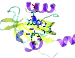
In this work we exploited time-resolved photoacoustics (PA) and molecular dynamics (MD) simulations to investigate the function of a conserved phenylalanine residue in blue sensing (BL) LOV domains. The LOV photocycle involves reversible formation of a photoproduct (LOV390) where the flavin mononucleotide (FMN) chromophore is covalently bound to a cysteine. LOV390 thermally returns to the dark adapted state (LOV447) with a lifetime τrec (s-to-h). In the LOV domain of Bacillus subtilis BsYtvA, the conserved F46 is one of the few residues undergoing a pronounced light-driven conformational change. PA and spectroscopic data show that in the YtvA variants F46A and F46Y light-induced structural changes are much smaller than those in the wild type (wt) protein, τrec is strongly accelerated and the energy content of LOV390 is lower for F46Y. MD simulations for each variant in the LOV447 and LOV390 states revealed an overall very stable structure of the BsYtvA-LOV domain. The largest variations emerged for the conserved HB network that includes FMN, Q123 (the “flipping” glutamine of LOV domains), and the conserved N104 and N94, with strong dependence on the presence of water. The lateral chain of Q123 in wt-LOV447 can adopt three alternative conformations, and movements act in concert with F46 flexibility. In LOV390, Q123 remains instead fixed in the orientation adopted in the crystal structure. Interestingly, in F46A, Q123 is locked in a LOV447-like conformation (pseudo-dark-adapted state), in both LOV447 and LOV390. In LOV447 of F46Y the tyrosine hydroxyl group fixes a water molecule, which induces a Q123 conformation similar to wt-LOV390, i.e. a pseudo-photoproduct state. These pseudo-dark-adapted and photoproduct-like conformations of the Q123 sidechain may account for the strong acceleration of the photocycle in the two variants. Given the importance of the “flipping” glutamine in light-to-signal propagation in LOV proteins, the results presented here underscore a crucial structural and functional role of the conserved F46. MD results also indicate that F46 is not directly engaged in permeability of the FMN pocket, but is involved in solvent ordering and the formation of water bridges.
- This article is part of the themed collection: Photofunctional flavoproteins and UVB photosensors
 Please wait while we load your content...