
Fluorescence characteristics of lipofuscin fluorophores from human retinal pigment epithelium

Abstract
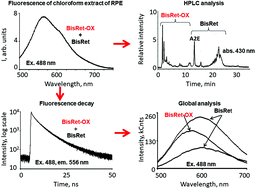
Lipofuscin granules accumulate in the retinal pigment epithelium (RPE) with age, especially in patients with visual diseases, including progressive age-related macular degeneration (AMD). Bisretinoids and their photooxidation and photodegradation products are major sources of lipofuscin granule fluorescence. The present study focused on examining the fluorescence decay characteristics of bisretinoid photooxidation and photodegradation products to evaluate the connection between fluorescence lifetime and spectral characteristics of target fluorophore groups. The primary objective of the study was to apply experimental spectral analysis results of lipofuscin granule fluorescence properties to interpretation of fluorescence lifetime imaging ophthalmoscopy data. Fluorescence analysis of the lipofuscin granule fluorophores in RPE collected from cadaver eyes was performed. The fluorescence lifetimes were measured by picosecond-resolved time correlated single photon counting technique. A global analytical method was applied to analyze data sets. The photooxidation and photodegradation products of bisretinoids exhibited a longer fluorescence lifetime (average value approximately 6 ns) and a shorter wavelength maximum (530–580 nm). Further, these products significantly contributed (more than 30%), to total fluorescence compared to the other fluorophores in lipofuscin granules. Thus, the contribution of oxidized lipofuscin bisretinoids to autofluorescence decay kinetics is an important characteristic for fluorescence lifetime imaging microscopy data analysis. The higher average fluorescence lifetime in AMD eyes was likely due to the higher abundance of oxidized bisretinoids compared with non-oxidized bisretinoids. Because higher level of oxidized bisretinoids is indicative of pathological processes in the retina and RPE, the present findings have the potential to improve fluorescence lifetime imaging approaches for early diagnosis of degenerative processes in the retina and RPE.
 Please wait while we load your content...