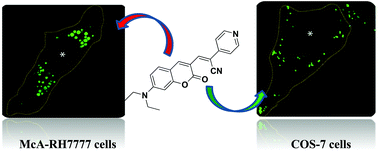
Live-cell imaging of lipid droplets using solvatochromic coumarin derivatives†
Abstract
Lipid droplets (LDs), the lipid-rich intracellular organelles, serve to regulate many physiological processes and therefore attention has been attracted towards their selective detection. We report positively solvatochromic lipophilic dyes, based on the push–pull framework containing coumarin–pyridine heterocycles for selective live-cell imaging of lipid droplets (LDs) in Cos-7 and McA-RH7777 cells at ultralow concentrations (200 nM). The fluorescent probes show a remarkable increase in fluorescence intensity with time with the hydrophobic core of the lipid droplets contributing to the observed intensity enhancement. The simple structural framework, red emission, strong Stokes shift (>80 nm), and excellent biocompatibility highlight their significance as a versatile imaging tool for studying lipid droplets (LDs).

- This article is part of the themed collection: Chemical Biology in OBC


 Please wait while we load your content...