
Selenium-deficient diet induces necroptosis in the pig brain by activating TNFR1 via mir-29a-3p†
 *ab
*ab
Abstract
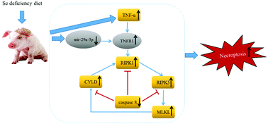
Selenium (Se) deficiency is one of the crucial factors related to nervous system disease and necroptosis. MicroRNAs (miRNAs) play vital roles in regulating necroptosis. However, the mechanism of Se deficiency-induced necroptosis in the pig brain tissue and the role that miRNAs play in this process are unclear. Therefore, in this study, in vitro and pig models of Se deficiency were replicated, and electron microscopy, quantitative real-time polymerase chain reaction (qRT-PCR) and western blot assays were performed. The results showed that brain cells typically undergo necrotic changes, and that Se deficiency suppresses mir-29a-3p, which increases the levels of TNFRSF1A (TNFR1). Subsequently, a distinct increase in the necroptosis markers (RIPK1, RIPK3, and MLKL) and an evident decrease in caspase 8 was observed. And the expression of 10 selenoproteins was decreased. Moreover, the in vitro experiments showed that the expression of mir-29a-3p decreased as the Se content in the medium decreased and the application of an mir-29a-3p inhibitor increased the number of necrotic cells and the accumulation of ROS, and these effects were inhibited by necrostatin-1 (Nec-1) and N-acetyl-cysteine (NAC), respectively. Taken together, we proved that Se deficiency induced necroptosis both in vitro and in vivo through the targeted regulation of TNFR1 by mir-29a-3p in the pig brain.
 Please wait while we load your content...