Issue 88, 2020

Direct visualization of the protein corona using carbon nanodots as a specific contrasting agent

Abstract

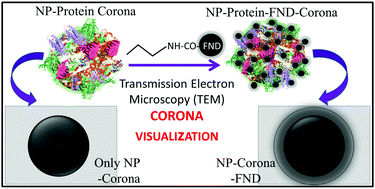
The cellular uptake of the nanoparticles is greatly affected by the formation of protein corona. As a result, an in-depth knowledge of direct visualization of the corona and quantification thereof is extremely important. Although transmission electron microscopy is one of the best techniques for visualization, the heavy metals that are used to increase the contrast of protein are non-specific and may lead to artifacts and erroneous conclusions. Here, we present a new strategy using carbogenic nanodots that showed excellent contrast, under a transmission electron microscope for the direct visualization and quantification of the single particle protein corona.

Graphical abstract: Direct visualization of the protein corona using carbon nanodots as a specific contrasting agent

Supplementary files

Article information

Article type
Communication
Submitted
20 Sep 2020
Accepted
06 Oct 2020
First published
06 Oct 2020

Chem. Commun., 2020,56, 13599-13602

Direct visualization of the protein corona using carbon nanodots as a specific contrasting agent

C. Rao, A. Yadav, R. Kaur, A. Prasad and C. K. Nandi, Chem. Commun., 2020, 56, 13599 DOI: 10.1039/D0CC06333A

To request permission to reproduce material from this article, please go to the Copyright Clearance Center request page.

If you are an author contributing to an RSC publication, you do not need to request permission provided correct acknowledgement is given.

If you are the author of this article, you do not need to request permission to reproduce figures and diagrams provided correct acknowledgement is given. If you want to reproduce the whole article in a third-party publication (excluding your thesis/dissertation for which permission is not required) please go to the Copyright Clearance Center request page.

Read more about how to correctly acknowledge RSC content.

Social activity

Spotlight

Advertisements