A colorful approach towards developing new nano-based imaging contrast agents for improved cancer detection†
Abstract
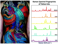
Providing physicians with new imaging agents to help detect cancer with better sensitivity and specificity has the potential to significantly improve patient outcomes. Development of new imaging agents could offer improved early cancer detection during routine screening or help surgeons identify tumor margins for surgical resection. In this study, we evaluate the optical properties of a colorful class of dyes and pigments that humans routinely encounter. The pigments are often used in tattoo inks and the dyes are FDA approved for the coloring of foods, drugs, and cosmetics. We characterized their absorption, fluorescence and Raman scattering properties in the hopes of identifying a new panel of dyes that offer exceptional imaging contrast. We found that some of these coloring agents, coined as “optical inks”, exhibit a multitude of useful optical properties, outperforming some of the clinically approved imaging dyes on the market. The best performing optical inks (Green 8 and Orange 16) were further incorporated into liposomal nanoparticles to assess their tumor targeting and optical imaging potential. Mouse xenograft models of colorectal, cervical and lymphoma tumors were used to evaluate the newly developed nano-based imaging contrast agents. After intravenous injection, fluorescence imaging revealed significant localization of the new “optical ink” liposomal nanoparticles in all three tumor models as opposed to their neighboring healthy tissues (p < 0.05). If further developed, these coloring agents could play important roles in the clinical setting. A more sensitive imaging contrast agent could enable earlier cancer detection or help guide surgical resection of tumors, both of which have been shown to significantly improve patient survival.

- This article is part of the themed collection: Biomaterials for Imaging and Sensing


 Please wait while we load your content...