Stabilization of indocyanine green dye in polymeric micelles for NIR-II fluorescence imaging and cancer treatment†
Abstract
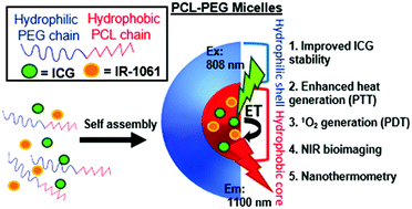
One of the most commonly used near infrared (NIR) dyes is indocyanine green (ICG), which has been extensively used for NIR bioimaging, photothermal and photodynamic therapy. However, upon excitation this dye can react with molecular oxygen to form singlet oxygen (SO), which can then cleave ICG to form non-fluorescent debris. In order to reduce the reaction between ICG and oxygen, we used energy transfer (ET) between the former and the NIR dye IR-1061. The two dyes were encapsulated in micelles composed of biocompatible poly(ethylene glycol)-block-poly(ε-caprolactone) (PCL–PEG). Micelles were characterized for their size using dynamic light scattering (DLS) and were found to measure about 35 nm in diameter. Fluorescence emission measurements were conducted to show that the stability of ICG against photodecomposition is increased. Moreover, this increased stability allows the encapsulated dye to generate more heat and for a longer time, compared to its free form. Studies with a SO indicator showed that as more IR-1061 is added to the micelles, less SO is produced. These results show how by changing the amount of added IR-1061 it is possible to tune the heat and SO generated by the system. Cell viability studies demonstrated that while particles were nontoxic under physiological conditions, upon 808 nm irradiation they become potent at eradicating MCF7 cancer cells. Moreover, it was demonstrated that both the increase of temperature and the creation of decomposition debris play a role in the cytotoxic efficacy of the micelles. Dye-loaded micelles that were injected to live mice showed bright fluorescence in the over 1000 nm NIR (OTN-NIR) region, allowing for visualization of blood vessels and internal organs. Most importantly, the encapsulated dyes remained stable for over 30 minutes, gradually accumulating in the liver and spleen. The presence of IR-1061 in addition to the heat-generating dye ICG allowed for simultaneous temperature modification and monitoring. We were able to assess the change in temperature by measuring the change in the fluorescence intensity of IR-1061 in the OTN-NIR region, a range with deep penetration of living tissues. These features illustrate the potential use of ICG/IR-1061 in PCL–PEG micelles as promising candidates for cancer treatment and diagnosis.


 Please wait while we load your content...