The effect of metal ions on endogenous melanin nanoparticles used as magnetic resonance imaging contrast agents†

Abstract
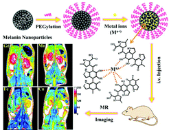
Melanin nanoparticles are of great importance in biomedicine. They have excellent affinity for metallic cations, especially paramagnetic ions, which has sparked interest in their application in the development of magnetic resonance imaging (MRI) contrast agents. In this work, we prepared ultrasmall water-soluble melanin nanoparticles, and investigated the binding properties of melanin toward different metal cations (Gd3+, Mn2+, Fe3+ and Cu2+), and compared their physicochemical properties and the MRI contrast enhancement ability in various metal chelated forms (MNP-PEG-M) in vitro and in vivo. We show that the saturation binding numbers of Gd3+, Mn2+, Fe3+ and Cu2+ per MNP-PEG were 49, 59, 69 and 62, respectively. MNP-PEG-Gd, MNP-PEG-Mn, MNP-PEG-Fe and MNP-PEG-Cu exhibited the maximum r1 relaxivities at the loading mass ratios of Gd3+ : MNP = 1 : 1, Mn2+ : MNP = 0.5 : 1, Fe3+ : MNP = 0.1 : 1 and Cu2+ : MNP = 0.1 : 1, corresponding to 49, 57, 54 and 51 chelated metals per MNP-PEG, respectively. The maximal per metal ion r1 relaxivity values were 61.9, 48.7, 11.1 and 9.7 mM−1 s−1 for MNP-PEG-Gd, MNP-PEG-Mn, MNP-PEG-Fe and MNP-PEG-Cu at 1.5 T, respectively. MNP-PEG-Gd and MNP-PEG-Fe presented larger sizes (6.9 nm and 5.8 nm) than MNP-PEG-Mn and MNP-PEG-Cu (3.4 nm and 3.7 nm), all featuring excellent solubility, high stability and ultrasmall size. A significant in vivo MRI signal enhancement in tissues was observed for all MNP-PEG-M after intravenous injection in mice, and these nanoparticles were excreted through renal and hepatobiliary pathways. In agreement with their r1 relaxivity values, MNP-PEG-Gd and MNP-PEG-Mn showed a significantly greater in vivo tissue maximum enhancement than MNP-PEG-Fe and MNP-PEG-Cu. This study could yield valuable insight into the development of a new class of MRI contrast agents.
 Please wait while we load your content...

