Microfluidic perfusion modulates growth and motor neuron differentiation of stem cell aggregates†
Abstract
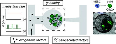
Microfluidic technologies provide many advantages for studying differentiation of three-dimensional (3D) stem cell aggregates, including the ability to control the culture microenvironment, isolate individual aggregates for longitudinal tracking, and perform imaging-based assays. However, applying microfluidics to studying mechanisms of stem cell differentiation requires an understanding of how microfluidic culture conditions impact cell phenotypes. Conventional cell culture techniques cannot directly be applied to the microscale, as microscale culture varies from macroscale culture in multiple aspects. Therefore, the objective of this work was to explore key parameters in microfluidic culture of 3D stem cell aggregates and to understand how these parameters influence stem cell behavior and differentiation. These studies were done in the context of differentiation of embryonic stem cells (ESCs) to motor neurons (MNs). We assessed how media exchange frequency modulates the biochemical microenvironment, including availability of exogenous factors (e.g. nutrients, small molecule additives) and cell-secreted molecules, and thereby impacts differentiation. The results of these studies provide guidance on how key characteristics of 3D cell cultures can be considered when designing microfluidic culture parameters. We demonstrate that discontinuous perfusion is effective at supporting stem cell aggregate growth. We find that there is a balance between the frequency of media exchange, which is needed to ensure that cells are not nutrient-limited, and the need to allow accumulation of cell-secreted factors to promote differentiation. Finally, we show how microfluidic device geometries can influence transport of biomolecules and potentially promote asymmetric spatial differentiation. These findings are instructive for future work in designing devices and experiments for culture of cell aggregates.


 Please wait while we load your content...