Lysosome-targeting pH indicator based on peri-fused naphthalene monoimide with superior stability for long term live cell imaging†
 ab
Javier
Garcia Lopez,
cd
Anela
Ivanova,
e
Kalina
Peneva
cd
and
Ute
Neugebauer
*abdef
ab
Javier
Garcia Lopez,
cd
Anela
Ivanova,
e
Kalina
Peneva
cd
and
Ute
Neugebauer
*abdef
Abstract
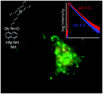
Lysosomes, the acidic degradation compartments of eukaryotic cells, play an essential role in many physiological processes. Their dysfunction is associated with a number of diseases, which are often related to an altered localization or luminal pH. Thus, the in-depth characterization of lysosomes within the intact eukaryotic cell is of utmost interest. For microscopic evaluation of lysosomal distribution and acidity, a number of labels have been developed, but many showed poor organelle specificity or rapid clearing from lysosomes, rendering them unsuitable for long-term observations. Here, we describe the synthesis and spectroscopic properties of a novel small molecule marker for lysosomes based on naphthalene monoimide with reversible, pH-dependent spectral shifts in both the absorption and the emission spectrum and acidity-associated changes in fluorescence lifetime. The dye can be excited either with single- or two-photon excitation and appears to be very stably associated with lysosomes for several days. We used this chromophore to detect chemically-induced changes of lysosomal pH in HeLa cells by ratiometric and FLIM imaging.
 Please wait while we load your content...

