Issue 48, 2019

Light-driven visualization of endogenous cysteine, homocysteine, and glutathione using a near-infrared fluorescent probe

Abstract

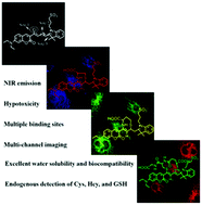
Herein, we presented a hydrosoluble triple-site and triple-excitation alternative NIR fluorescent probe for visualization of endogenous biothiols in phosphate-buffered saline (pH 7.4, 10 mM). Upon irradiation using different excitation light, probe 1 exhibited different fluorescence responses upon the addition of Cys, Hcy, and GSH: λex = 419 nm, λem = 498 nm; λex = 518 nm, λem = 573, 616, 727, and 783 nm; λex = 555 nm, λem = 612 and 727 nm, respectively. Furthermore, 1 was favourably applied for bioimaging endogenous Cys, Hcy, and GSH in A375 cells through well-defined blue–green–red emission channels.

Graphical abstract: Light-driven visualization of endogenous cysteine, homocysteine, and glutathione using a near-infrared fluorescent probe

Supplementary files

Article information

Article type
Paper
Submitted
04 Aug 2019
Accepted
02 Nov 2019
First published
05 Nov 2019

J. Mater. Chem. B, 2019,7, 7723-7728

Light-driven visualization of endogenous cysteine, homocysteine, and glutathione using a near-infrared fluorescent probe

Y. Yang, Y. Wang, Y. Feng, C. Cao, X. Song, G. Zhang and W. Liu, J. Mater. Chem. B, 2019, 7, 7723 DOI: 10.1039/C9TB01645G

To request permission to reproduce material from this article, please go to the Copyright Clearance Center request page.

If you are an author contributing to an RSC publication, you do not need to request permission provided correct acknowledgement is given.

If you are the author of this article, you do not need to request permission to reproduce figures and diagrams provided correct acknowledgement is given. If you want to reproduce the whole article in a third-party publication (excluding your thesis/dissertation for which permission is not required) please go to the Copyright Clearance Center request page.

Read more about how to correctly acknowledge RSC content.

Social activity

Spotlight

Advertisements