Deciphering of cerebrovasculatures via ICG-assisted NIR-II fluorescence microscopy†

Abstract
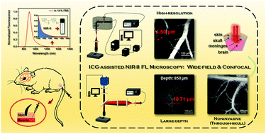
Benefiting from high spatial resolution and large penetration depth, NIR-II (second near-infrared spectral region, 900–1700 nm) fluorescence imaging based on US Food and Drug Administration (FDA)-approved indocyanine green (ICG) is expected to be a good approach for clinical applications. As of now, nearly all reported works on ICG-assisted NIR-II fluorescence imaging are macro-imaging while micro-angiography is also a significant imaging modality, especially during the diagnosis and treatment of cerebrovascular diseases. Herein, based on NIR-II fluorescence wide-field microscopy, the high-resolution observation of cerebral vasculature was performed at deep brain tissues in mice via intramuscular (IM) injection of ICG. Altered cerebral vessels in mice after brain embolism were further noticed by means of noninvasive through-skull NIR-II fluorescence microscopy. Moreover, ICG-assisted NIR-II fluorescence confocal microscopy was executed to observe cerebral vasculature, presenting optical sectioning capability and higher spatial resolution.
 Please wait while we load your content...

