Effects of hydroxyapatite surface nano/micro-structure on osteoclast formation and activity†
Abstract
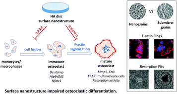
The surface structure of calcium phosphate (CaP) ceramic plays an important role in its osteoinductivity; however, little is known about its effects on osteoclastogenesis. In this study, an intramuscular implantation model suggested a potential relationship between hydroxyapatite (HA)-induced bone formation and osteoclast appearance in the non-osseous site, which might be modulated by scaffold surface structure. Then, three dense HA discs with different grain sizes from biomimetic nanoscale (∼100 nm) to submicron scale (∼500 nm) were fabricated via distinct sintering procedures, and their impacts on osteoclastic differentiation of RAW 264.7 macrophages under RANKL stimulation were further investigated. Our results showed that compared with the ones in the submicron-scale dimension, nano-structured HA discs markedly impaired osteoclastic formation and function, as evidenced by inhibited cell fusion, reduced osteoclast size, less-defined actin ring, increased osteoclast apoptosis, suppressed expression of osteoclast specific genes and proteins, decreased TRAP-positive cells, and hampered resorption activity. This demonstrated that the surface structure of CaP ceramics has a great influence on osteoclastogenesis, which might be further related to its osteoinductive capacity. These findings might not only help us gain insight into biomolecular events during CaP-involved osteoinduction, but also offer a principle for designing orthopaedic implants with an ability of regulating both osteogenesis and osteoclastogenesis to achieve the desired performance.


 Please wait while we load your content...