
A skin-inspired 3D bilayer scaffold enhances granulation tissue formation and anti-infection for diabetic wound healing
 *b
and
*b
and
Abstract
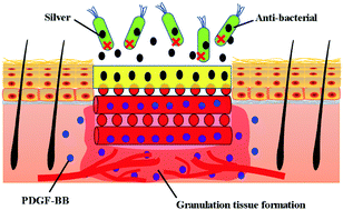
Diabetic wounds are characterized by sustained chronic inflammation, and decreased granulation tissue formation and vascularization, and it is a great challenge for researchers to promote chronic wound healing. In this study, we fabricate a bilayer skin substrate composed of a top layer made of silver-loaded gelatine cryogel and a bottom layer made of a platelet-derived growth factor-BB (PDGF-BB) loaded 3D printed gelatine scaffold for diabetic wound healing. In vitro, the concentration of loaded silver has no effect on the proliferation of fibroblasts, keratinocytes and immune cells cocultured with silver-loaded 3D bilayer hydrogel extract medium. In addition, the CFU quantification results showed that the release of silver nanoparticles was able to significantly kill bacteria including Pseudomonas aeruginosa, Staphylococcus aureus and Escherichia coli. In vivo, PDGF-BB-loaded scaffolds, and silver and PDGF-BB coloaded scaffolds were able to accelerate wound closure, re-epithelialization, granulation tissue formation and angiogenesis compared to the scaffold and silver-loaded scaffold groups at each indicated time point. The silver and PDGF-BB coloaded bilayer hydrogel scaffolds show great potential for promoting diabetic wound healing and against bacterial infections.
 Please wait while we load your content...

