Investigation of the magnetosome biomineralization in magnetotactic bacteria using graphene liquid cell – transmission electron microscopy
Abstract
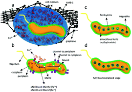
Understanding the biomineralization pathways in living biological species is a grand challenge owing to the difficulties in monitoring the mineralization process at sub-nanometer scales. Here, we monitored the nucleation and growth of magnetosome nanoparticles in bacteria and in real time using a transmission electron microscope (TEM). To enable biomineralization within the bacteria, we subcultured magnetotactic bacteria grown in iron-depleted medium and then mixed them with iron-rich medium within graphene liquid cells (GLCs) right before imaging the bacteria under the microscope. Using in situ electron energy loss spectroscopy (EELS), the oxidation state of iron in the biomineralized magnetosome was analysed to be magnetite with trace amount of hematite. The increase of mass density of biomineralized magnetosomes as a function of incubation time indicated that the bacteria maintained their functionality during the in situ TEM imaging. Our results underpin that GLCs enables a new platform to observe biomineralization events in living biological species at unprecedented spatial resolution. Understanding the biomineralization processes in living organisms facilitates the design of biomimetic materials, and will enable a paradigm shift in understanding the evolution of biological species.


 Please wait while we load your content...