
Aggregation-induced emission based PET probe for liver function imaging†
 *a
*a
Abstract
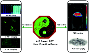
To locate the site of a liver lesion by positron emission tomography (PET) imaging and then remove the lesion site under the guidance of fluorescence imaging, we designed an aggregate-induced emission (AIE)-based PET probe [nat/68Ga] 5 based on the effects of the reticuloendothelial system. By modifying derivatives of tetraphenylethene, we obtained a bifunctional chelating agent with AIE characteristics, which can complex with natGa3+ or 68Ga3+ at room temperature to form [natGa] 5 or [68Ga] 5. [natGa] 5 has typical AIE characteristics and can form nanomicelles when its concentration is greater than the critical micelle concentration (CMC). Cellular fluorescence confocal imaging experiments and radioactive uptake experiments showed that Kupffer cells had a high uptake of [nat/68Ga] 5 while HepG2 cells had lower uptake of [nat/68Ga] 5, indicating that liver function imaging was achieved by the phagocytosis of [nat/68Ga] 5 by Kupffer cells into the liver. A biodistribution study showed that [68Ga] 5 shows high accumulation in the liver after intravenous injection into rats. The in vivo PET imaging profile demonstrated an excellent liver to muscle ratio. Thus, [nat/68Ga] 5 could potentially be used as a liver function PET imaging agent for clinical diagnosis and intraoperative navigation.
 Please wait while we load your content...

