3D spheroids generated on carbon nanotube-functionalized fibrous scaffolds for drug metabolism and toxicity screening
 *a
*a
Abstract
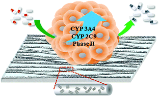
The mechanical and electrical stimuli have a profound effect on the cellular behavior and function. In this study, a series of conductive nanofibrous scaffolds are developed by blend electrospinning of poly(styrene-co-maleic acid) (PSMA) and multiwalled-carbon nanotubes (CNTs), followed by grafting galactose as cell adhesion cues. When the mass ratios of CNTs to PSMA increase up to 5%, the alignment, Young's modulus and conductivity of fibrous scaffolds increase, whereas the average diameter, pore size and elongation at break decrease. Primary hepatocytes cultured on the scaffolds are self-assembled into 3D spheroids, which restores the hepatocyte polarity and sufficient expression of drug metabolism enzymes over an extended period of time. Among these conductive scaffolds, hepatocytes cultured on fibers containing 3% of CNTs (F3) show the highest clearance rates of model drugs, offering a better prediction of the in vivo data with a high correlation value. Moreover, the drug metabolism capability is maintained over 15 days and is more sensitive towards the inducers and inhibitors of metabolizing enzymes, demonstrating the applicability for drug–drug interaction studies. Thus, this culture system has been demonstrated as a reliable in vitro model for high-throughput screening of metabolism and toxicity in the early phases of drug development.
 Please wait while we load your content...

