Identification of early inflammatory changes in the tympanic membrane with Raman spectroscopy
 a
a
Abstract
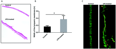
The tympanic membrane (TM) is a dynamic structure that separates the middle ear from the external auditory canal. It is also integral for the transmission of sound waves. In this study, we demonstrate the feasibility of using Raman spectroscopy to identify early chemical changes resulting from inflammation in the TM that can serve as an indicator of acute otitis media. Bacterial lipopolysaccharide (LPS) was injected trans-tympanicaly in a murine model. Presence of inflammatory response was assessed with binocular microscopy, confirmed with histopathology and immunofluorescence staining. Successful discrimination suggesting spectral differences among the control and LPS treated groups was achieved using principal component analysis. Raman imaging revealed major differences in collagen distribution and nucleic acid content. Image segmentation analysis on the trichrome stained tissue sections was performed to corroborate the Raman spectra. The spectral co-localization study suggests changes in the expression of collagen IV specific signals in LPS treated samples. The overall findings of the study support prospective application of RS in the diagnosis and therapeutic monitoring of otitis media.
 Please wait while we load your content...

