
Yolk–shell structured Au@Ag@mSiO2 as a probe for sensing cysteine enantiomers and Cu2+ based on circular dichroism†
 *a
*a
Abstract
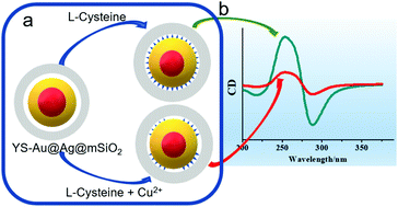
A yolk–shell structured Au@Ag@mSiO2 probe was fabricated by coating a layer of mesoporous silica on the surface of a Au@Ag core–shell nanosphere, followed by partially removing the Ag shell. Mirror symmetrical chiral signals at ∼258 nm in the UV region were observed for the probe upon coupling with cysteine enantiomers on the surface of Au@Ag. The intensity of the CD signal of the probe is enhanced by increasing L/D-Cys concentration, allowing the quantitative determination of the cysteine enantiomers. The developed method shows an excellent linear relationship between the CD signal and L-Cys concentration ranging from 10 μM to 90 μM with a limit of detection of 8.5 μM. In the presence of Cu2+, the CD signal of the probe weakened due to the oxidation of L-cysteine to L-cystine catalyzed by Cu2+. Based on this phenomenon, a new strategy for the detection of Cu2+ can be developed. Under the optimized conditions, the CD signal decreases linearly with the log of the concentration of Cu2+ in the range from 1 to 250 nM with a detection limit of 0.1 nM.
 Please wait while we load your content...

