
The imaging of local glucose levels in tumor periphery via peroxyoxalate chemiluminescent nanoparticle–glucose oxidase–doped alginate hydrogel†
 a
and
Wei
Tang
*a
a
and
Wei
Tang
*a
Abstract
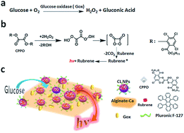
Glucose is a metabolic intermediate of living cells. Tumor cells exploit anaerobic glycolysis for their energy needs and cause a greater glucose consumption than that is observed in normal tissue. Therefore, the detection and imaging of glucose levels in tumor tissues serves as an effective method for the detection and staging of tumors. Peroxyoxalate chemiluminescence (POCL) shows high sensitivity and selectivity toward hydrogen peroxide (H2O2), which offers a high imaging specificity for H2O2 and glucose in tumor, as glucose can react with the dissolved oxygen to produce H2O2 in the presence of glucose oxidase (GOx). Herein, we reported a simple POCL–GOx–alginate/Ca gel system to detect and image the local concentration of glucose in the tumor periphery. We constructed nanoparticles enwrapping rubrene and CPPO as a CL nanoreactor and mixed with GOx. Then, alginic acid sodium salt solution was added to form a POCL–GOx–alginate solution. When Ca2+ was added into this solution, alginate could gelate to render the POCL-NPs/GOx to gather around the tumor and prevent the nanoparticles from dispersing. When glucose permeated into this POCL–GOx–alginate/Ca gel system, it was oxidized by GOx and H2O2 was produced. Then, H2O2 ignited the CPPO/rubrene nanoreactor, and the CL intensity was observed immediately. Our results showed that this POCL–GOx–alginate/Ca gel system could quantitatively detect glucose in vitro and successfully be applied for the in vivo imaging of the local glucose concentration in a tumor-bearing mouse. Our strategy had a high signal-to-noise ratio because of the high efficiency of the oxidation of GOx due to the abundant supply of oxygen in tumor periphery and the gathering of CL reactors by alginate/Ca gel network. Owing to the high biological safety of alginate, this simple and low-cost CL/gel system has a potential to be used as a partial alternate for FDG-PET in clinical diagnose. Furthermore, this CL nanoparticle–oxidase enzyme–alginate/Ca gel system provides a platform for the in situ imaging of substrates of other oxidase enzymes in vivo.
- This article is part of the themed collection: Analytical Methods Recent HOT articles
 Please wait while we load your content...

