Issue 10, 2018

Multicolor monitoring of cellular organelles by single wavelength excitation to visualize the mitophagy process

Abstract

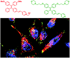
Multiplexed cellular organelle imaging using single wavelength excitation is highly desirable for unravelling cellular functions but remains challenging. This requires the design of organelle specific fluorophores with distinct emission but similar absorption. Herein, we present two unique aggregation-induced emission (AIE) probes to track mitochondria and lysosomes simultaneously with emission colors that can be distinguished from that of the nucleus stain Hoechst 33342 upon single wavelength excitation. Compared to conventional organelle stains, the two AIE probes have larger Stokes shifts and higher photostability, which endow them with the capability to monitor bioprocesses, such as mitophagy with strong and sustained fluorescent signals. Moreover, both probes can also stain intracellular organelles in zebrafish larvae with good cell-penetrating capabilities, showing their great potential to monitor bioprocesses in vivo.

Graphical abstract: Multicolor monitoring of cellular organelles by single wavelength excitation to visualize the mitophagy process

Supplementary files

Article information

Article type
Edge Article
Submitted
23 Oct 2017
Accepted
01 Feb 2018
First published
02 Feb 2018
This article is Open Access

All publication charges for this article have been paid for by the Royal Society of Chemistry
Creative Commons BY-NC license

Chem. Sci., 2018,9, 2756-2761

Multicolor monitoring of cellular organelles by single wavelength excitation to visualize the mitophagy process

F. Hu, X. Cai, P. N. Manghnani, Kenry, W. Wu and B. Liu, Chem. Sci., 2018, 9, 2756 DOI: 10.1039/C7SC04585A

This article is licensed under a Creative Commons Attribution-NonCommercial 3.0 Unported Licence. You can use material from this article in other publications, without requesting further permission from the RSC, provided that the correct acknowledgement is given and it is not used for commercial purposes.

To request permission to reproduce material from this article in a commercial publication, please go to the Copyright Clearance Center request page.

If you are an author contributing to an RSC publication, you do not need to request permission provided correct acknowledgement is given.

If you are the author of this article, you do not need to request permission to reproduce figures and diagrams provided correct acknowledgement is given. If you want to reproduce the whole article in a third-party commercial publication (excluding your thesis/dissertation for which permission is not required) please go to the Copyright Clearance Center request page.

Read more about how to correctly acknowledge RSC content.

Social activity

Spotlight

Advertisements