Distribution of superparamagnetic Au/Fe nanoparticles in an isolated guinea pig brain with an intact blood brain barrier†
 ‡a
Galina V.
Beznoussenko,
d
Alexander A.
Mironov,
d
Carolina
Frassoni,
c
Francesco
Stellacci,
ef
Marco
de Curtis
b
and
Silke
Krol
*ag
‡a
Galina V.
Beznoussenko,
d
Alexander A.
Mironov,
d
Carolina
Frassoni,
c
Francesco
Stellacci,
ef
Marco
de Curtis
b
and
Silke
Krol
*ag
Abstract
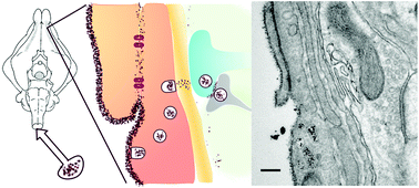
Diagnosis and treatment of brain disorders, such as epilepsy, neurodegenerative diseases and tumors, would benefit from innovative approaches to deliver therapeutic or diagnostic compounds into the brain parenchyma, with either a homogeneous or a targeted localized distribution pattern. To assess the mechanistic aspect of penetration of nanoparticles (NPs) into the brain parenchyma, a complex, yet controlled and facilitated environment was used: the isolated guinea pig brain maintained in vitro by arterial perfusion. In this unique preparation the blood–brain barrier and the interactions between vascular and neuronal compartments are morphologically and functionally preserved. In this study, superparamagnetic Au/Fe nanoparticles (MUS:OT Au/Fe NPs), recently studied as a promising magnetic resonance T2 contrast agent with high cellular penetration, were arterially perfused into the in vitro isolated brain and showed high and homogeneous penetration through transcytosis into the brain parenchyma. Ultramicroscopy investigation of the in vitro isolated brain sections by TEM analysis of the electron-dense core of the MUS:OT Au/Fe NPs was conducted to understand NPs’ brain penetration through the BBB after in vitro arterial perfusion and their distribution in the parenchyma. Our data suggest that MUS:OT Au/Fe NPs enter the brain utilizing a physiological route and therefore can be exploited as brain penetrating nanomaterials with potential contrast agent and theranostics capabilities.
 Please wait while we load your content...

