Fe3+-Enhanced NIR-to-NIR upconversion nanocrystals for tumor-targeted trimodal bioimaging†
Abstract
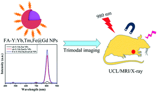
Multimodal bioimaging, which integrates the merits of two or more imaging techniques, has been applied in clinical prognosis and diagnosis for several years. For in vivo bioimaging, it is important to develop materials with excitation and emission spectra that lie in the “optical transparent biological window” (750–1000 nm), in which there is minimal autofluorescence, little risk of damage to biosamples and deep tissue penetration ability. Herein, folate conjugated core–shell upconversion luminescence (UCL) nanoparticles (NPs), FA-NaYF4:Yb3+,Tm3+,20%Fe3+@NaGdF4 (FA-Y:Yb,Tm,Fe@Gd) NPs, were synthesized and applied for trimodal imaging, i.e. NIR UCL imaging using the Y:Yb,Tm,Fe core, and magnetic resonance and X-ray imaging using the NaGdF4 shell. The folate was incorporated to be used as a tumor-targeting probe. The addition of 20%Fe3+ (in moles) resulted in a 20 times increase in the NIR UCL intensity under 980 nm excitation. The role of the Fe3+ in the enhancement of the NIR UCL efficiency was explored. The methyl thiazolyl tetrazolium (MTT) assay of HeLa cells and the histological analysis of healthy viscera sections showed that the NPs have low biological toxicity.


 Please wait while we load your content...