Cytotoxicity studies of selected cadmium-based quantum dots on 2D vs. 3D cell cultures
Abstract
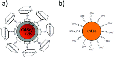
In our work, the cytotoxicity of selected, cadmium-based quantum dots with various surface architectures was studied on 3D spheroids. A specially designed microsystem as a tool for three-dimensional cell culture and nanoparticle toxicity evaluation was used for this aim. Two types of hydrophilic quantum dots with different surface charges at physiological pH were examined: CdTe-capped with ω-mercaptocarboxylic acid (COO−-terminated) and CdSeS/ZnS-glucosamine (–OH-terminated). We studied the influence of five different concentrations of nanoparticles within the range of 5 to 100 μM in order to assess dose-dependent toxic effects of the selected quantum dots on cellular spheroids as a more realistic (in vivo-like) model of human tissue. The obtained results of cytotoxicity were compared with the results for a standard, two-dimensional model – a cell monolayer. In the case of three-dimensional structures of various cell lines (normal MRC-5 and tumor A549) no significant differences in cytotoxicity caused by the tested nanoparticles have been noticed. The comparative studies revealed the enhanced biocompatibility of CdSeS/ZnS-OH quantum dots resulting from the presence of an uncharged ligand of biomimetic character on their surface. It was also found that the cytotoxicity of the same quantum dots depends on the cell culture model on which the tests were conducted. Significantly higher cytotoxicity of the tested nanomaterial was observed when experiments were carried out with the use of cell monolayers. Based on the obtained results, we claim that cytotoxicity studies of nanomaterials conducted on standard 2D cell monolayers are overestimated. In our opinion, reliable in vitro studies on the biological activity of nanoparticles require application of 3D cell cultures.


 Please wait while we load your content...