
Transition metals and trace elements in the retinal pigment epithelium and choroid: correlative ultrastructural and chemical analysis by analytical electron microscopy and nano-secondary ion mass spectrometry†
 *a
*a
Abstract
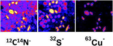
Understanding the localisation and abundance of structural elements, trace elements and especially transition metals like Cu and Zn in ocular tissue sections is important for physiology, and also for the characterisation of diseases related to oxidative stress like age-related macular degeneration. Transition metal abundances were investigated in an aged donor eye by nano-secondary ion mass spectrometry (nano-SIMS) elemental mapping using Cs+ and O− primary ions, respectively, and correlated to their respective mole fractions investigated by analytical electron microscopy (AEM). The ultrastructure of the tissue and the elemental composition of melanosomes of the choroid and RPE, and RPE lipofuscin and melanolipofuscin granules can adequately be investigated by nano-SIMS using the secondary ion maps. Melanosomes, 0.5–1 μm in size, yield sulphur maps and maps of stored metals like calcium, sodium and copper. Lipofuscin shows especially high phosphorus signals. Elements with mole fractions of about 0.1 at%, e.g. for P and Cu, as investigated by AEM before, can be validated using simultaneous SIMS maps with an estimated lateral resolution of 66 nm with typical acquisition times of 30 minutes for each area of interest. However, Zn (0.19 at%) was not detected by SIMS. Nano-SIMS imaging of CN−, PO2−, S−, Cu−, Ca+, Fe+ and Na+ ions provides excellent detection limits demonstrating the possibilities for chemical mapping with high-sensitivity trace element detection and reduced acquisition times. Quantification of nano-SIMS data was achieved by correlating mole fractions obtained by AEM to secondary ions per pixel obtained by nano-SIMS. Both methods yield the melanin type in melanosomes and trace metal storage.
 Please wait while we load your content...