A three-dimensional in vitro dynamic micro-tissue model of cardiac scar formation†
Abstract
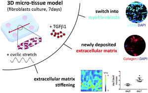
In vitro cardiac models able to mimic the fibrotic process are paramount to develop an effective anti-fibrosis therapy that can regulate fibroblast behaviour upon myocardial injury. In previously developed in vitro models, typical fibrosis features were induced by using scar-like stiffness substrates and/or potent morphogen supplementation in monolayer cultures. In our model, we aimed to mimic in vitro a fibrosis-like environment by applying cyclic stretching of cardiac fibroblasts embedded in three-dimensional fibrin-hydrogels alone. Using a microfluidic device capable of delivering controlled cyclic mechanical stretching (10% strain at 1 Hz), some of the main fibrosis hallmarks were successfully reproduced in 7 days. Cyclic strain indeed increased cell proliferation, extracellular matrix (ECM) deposition (e.g. type-I-collagen, fibronectin) and its stiffness, forming a scar-like tissue with superior quality compared to the supplementation of TGFβ1 alone. Taken together, the observed findings resemble some of the key steps in the formation of a scar: (i) early fibroblast proliferation, (ii) later phenotype switch into myofibroblasts, (iii) ECM deposition and (iv) stiffening. This in vitro scar-on-a-chip model represents a big step forward to investigate the early mechanisms possibly leading later to fibrosis without any possible confounding supplementation of exogenous potent morphogens.


 Please wait while we load your content...