Comparison of quenching mechanisms in Gd3Al5−xGaxO12:Ce3+ (x = 3 and 5) garnet phosphors by photocurrent excitation spectroscopy
Abstract
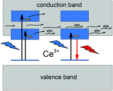
In this work we present the results of photocurrent excitation spectroscopy (PCE) of Gd3Al2Ga3O12:Ce3+ (GAGG:Ce3+) and Gd3Ga5O12:Ce3+ (GGG:Ce3+) performed at temperatures ranging from 100 to 500 K supplemented by spectroscopic measurements (steady state and time resolved photoluminescence spectroscopy) performed at temperatures ranging from 10 to 500 K and at high pressure up to 300 kbar. The PCE spectra contain bands related to transitions from the ground state 2F5/2 of the 4f1 electronic configuration to the crystal field split states related to the 5d1 electronic configuration of Ce3+. This implicates the presence of the autoionization process – transfer of electrons from the localized, excited states of Ce3+ to the conduction band (CB), directly linked to luminescence quenching of Ce3+. The mechanism of autoionization of GAGG:Ce3+ and GGG:Ce3+ was determined to be different on the grounds of differences in temperature dependence of photocurrent intensity. The latter system exhibits autoionization, which occurs when all of the 5d excited states are degenerated with the CB, whereas in the former system, the autoionization process is thermally assisted with an activation energy barrier (distance to the edge of the CB) of approximately 1600 cm−1. In GGG:Ce3+ the degeneracy of 5d1 states of Ce3+ was lifted by application of high pressure, shifting the edge of the CB up and exposing Ce3+ luminescence at 20 kbar. Further spectroscopic analyses of the pressure–temperature dependence of the luminescence decay time as well as the temperature dependence of photocurrent intensity of GGG:Ce3+ have independently shown existence of a luminescence quenching state located approximately 600 cm−1 below the CB, attributed to the impurity trapped exciton.


 Please wait while we load your content...