Issue 18, 2018

SEM–Raman image cytometry of cells

Abstract

Correlative and integrated scanning electron microscopy (SEM) and Raman micro-spectroscopy is presented that enables the characterization and identification of different cancer and non-cancer cells through SEM–Raman image cytometry. The hybrid microscopy system enables the acquisition of high resolution SEM images of uncoated cells and the spatial correlation with chemical information as obtained from Raman micro-spectroscopic imaging. A sample preparation protocol and a workflow are presented that are compatible with the demands of hybrid SEM–Raman microscopy. Stainless steel cell substrates were used that are both conductive and give a low optical response in Raman scattering. Correlative and integrated SEM–Raman micro-spectroscopy is illustrated with cells from blood and cells from a SKBR-3 breast cancer cell line.
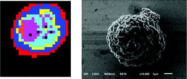
Graphical abstract: SEM–Raman image cytometry of cells

Article information

Article type
Paper
Submitted
23 May 2018
Accepted
30 Jul 2018
First published
02 Aug 2018

Analyst, 2018,143, 4495-4502

SEM–Raman image cytometry of cells

A. Enciso-Martinez, F. J. Timmermans, A. Nanou, L. W. M. M. Terstappen and C. Otto, Analyst, 2018, 143, 4495 DOI: 10.1039/C8AN00955D

To request permission to reproduce material from this article, please go to the Copyright Clearance Center request page.

If you are an author contributing to an RSC publication, you do not need to request permission provided correct acknowledgement is given.

If you are the author of this article, you do not need to request permission to reproduce figures and diagrams provided correct acknowledgement is given. If you want to reproduce the whole article in a third-party publication (excluding your thesis/dissertation for which permission is not required) please go to the Copyright Clearance Center request page.

Read more about how to correctly acknowledge RSC content.

Social activity

Spotlight

Advertisements