Identification and characterization of different tissues in blood vessel by multiplexed fluorescence lifetimes†

Abstract
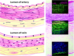
Herein, fluorescence lifetime imaging microscopy (FLIM) was used to directly measure eosin fluorescence lifetimes from H&E-stained umbilical artery, and a further utilization of eosin for high-content and multi-target analysis was proposed for the first time. Smooth muscles, collagens, and elastic fibers can be distinguished by eosin fluorescence lifetimes (P < 0.001). Erythrocytes, smooth muscles, elastic fibers, and type I and III collagen from the H&E-stained umbilical artery can be simultaneously identified by multiplexed fluorescence lifetimes of eosin. Use of eosin and lifetime-based separation is a potential method to simplify the special staining for clinicopathologic examination. Multiplexed eosin fluorescence lifetimes may be a newly developed method that can directly determine the relative content of elastic fiber and collagens from the H&E-stained sections. FLIM may have potential applications as an assisted tool in the assessment of the severity and complexity of cardiovascular diseases.
 Please wait while we load your content...

