Issue 3, 2018

Direct detection of lysine side chain NH3+ in protein–heparin complexes using NMR spectroscopy

Abstract

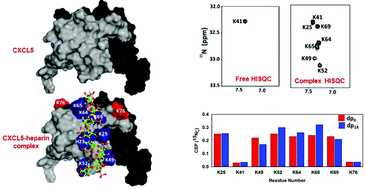
Two NMR observables, the NζH3+ peak in the HISQC spectrum and Nζ chemical shift difference between the free and heparin-bound forms, can identify binding-interface lysines in protein–heparin complexes. Unlike backbone chemical shifts, these direct probes are stringent and are less prone to either false positives or false negatives.

Graphical abstract: Direct detection of lysine side chain NH3+ in protein–heparin complexes using NMR spectroscopy

Article information

Article type
Communication
Submitted
24 Aug 2017
Accepted
13 Dec 2017
First published
13 Dec 2017

Analyst, 2018,143, 635-638

Direct detection of lysine side chain NH3+ in protein–heparin complexes using NMR spectroscopy

K. M. Sepuru, J. Iwahara and K. Rajarathnam, Analyst, 2018, 143, 635 DOI: 10.1039/C7AN01406F

To request permission to reproduce material from this article, please go to the Copyright Clearance Center request page.

If you are an author contributing to an RSC publication, you do not need to request permission provided correct acknowledgement is given.

If you are the author of this article, you do not need to request permission to reproduce figures and diagrams provided correct acknowledgement is given. If you want to reproduce the whole article in a third-party publication (excluding your thesis/dissertation for which permission is not required) please go to the Copyright Clearance Center request page.

Read more about how to correctly acknowledge RSC content.

Social activity

Spotlight

Advertisements