Synergistically enhanced upconversion luminescence in Li+-doped core–shell-structured ultrasmall nanoprobes for dual-mode deep tissue fluorescence/CT imaging†
Abstract
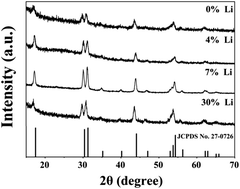
The development of upconversion luminescence that allows for multimodal imaging in terms of resolution and penetration depth using a single system is attracting increasing interest for use in clinical molecular imaging and diagnostics. In this study, a simple method for inducing high-intensity upconversion luminescence by doping Li+ ions in a core–shell-structured NaLuF4:Yb,Tm system was developed. The synergistic effects of Li+ doping and the shell layer enhanced the luminescence intensity by approximately 210 times. In vitro and in vivo experiments showed that the high-intensity luminescence of nanoparticles exhibited a depth penetration ability for biological tissue. Owing to the heavy atom effect of the Lu3+ ions, the nanoparticles, which had a size of 23 nm, showed good CT imaging performance when compared with a clinical contrast agent, in addition to allowing for deep tissue imaging. The excellent optical and CT imaging properties of the Li+-doped high-luminescence core–shell upconversion nanoparticles suggest that they are highly suited for use in both deep tissue fluorescence imaging and CT imaging for multimodal diagnosis.


 Please wait while we load your content...