Polymorphic light eruption and IL-1 family members: any difference with allergic contact dermatitis?
Abstract
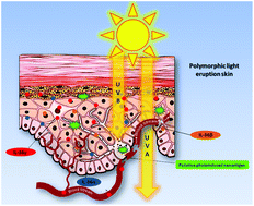
Polymorphic light eruption (PLE) is described as a delayed-type hypersensitivity reaction (DTHR) toward a de novo light-induced antigen, yet to be identified. In effect, the inflammatory pathways of PLE and allergic contact dermatitis (ACD) share common patterns in terms of the mediators involved from the innate and adaptive immune system participating in the DTHR. As we have previously highlighted the role of interleukin (IL)-1 family members in ACD, we hypothesised that the same mediators could have similar functions in PLE. Our research aimed to assess the expression of certain IL-1family members in PLE patients vs. controls, and to compare it with ACD. The study population comprised 17 patients with PLE, 5 affected by ACD and 10 healthy controls in the same age range. Lesional and healthy skin samples were collected respectively from patients and donors. IL-36α, IL-36β, IL-36γ, IL-36 receptor antagonist (Ra), IL-1β, IL-33 gene and protein expressions were evaluated through RT-PCR and immunohistochemistry. Circulating proteins in the PLE patients were analysed by using western blot. The IL-36γ gene expression was significantly increased in PLE lesions compared to that in healthy controls and ACD lesions (***p < 0.001; ##p < 0.01 respectively), whereas the other analyzed ILs were more expressed in ACD. Immunohistochemical analysis revealed that IL-36α and IL-36γ protein levels were increased in PLE lesions compared to those of the healthy samples (***p < 0.001). Furthermore the IL-36γ plasma level was increased in PLE patients vs. controls (*p < 0.05). Our findings indicate that the IL-1 family pro-inflammatory members are increased in PLE with distinct differences from those in ACD, in particular with regard to IL-36γ mRNA regulation. Their role as activators of the local, and perhaps systemic, immune response, or as inhibitors of the immune tolerance machinery, needs further investigation.


 Please wait while we load your content...