Surface-enhanced hyper Raman hyperspectral imaging and probing in animal cells†
Abstract
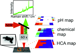
Hyper Raman scattering, that is, spontaneous, two-photon excited Raman scattering, of organic molecules becomes strong when it occurs as surface-enhanced hyper Raman scattering (SEHRS), in the proximity of plasmonic nanostructures. Its advantages over one-photon excited surface-enhanced Raman scattering (SERS) include complementary vibrational information resulting from different selection rules, probing of very small focal volumes, and beneficial excitation with long wavelengths. Here, imaging of macrophage cells by SEHRS is demonstrated, using SEHRS labels consisting of silver nanoparticles and two different molecules, 2-naphthalenethiol and para-mercaptobenzoic acid, that are excited off-resonance. The vibrational signatures of the molecules are discriminated using hyperspectral analysis and provide information about the subcellular localization of the SEHRS probes. The SEHRS based hyperspectral imaging approach presented here uses principal component analysis (PCA) to localize the reporter molecules inside the cells and is augmented by hierarchical cluster analysis (HCA). The high sensitivity of SEHRS spectra with respect to small environmental changes can be utilized for mapping of physiological parameters in the endosomal system of the cells. This is illustrated by discussing the spatial distribution of endosomes of varying pH inside the cytosol.


 Please wait while we load your content...