
Metal maps of sclerotic hippocampi of patients with mesial temporal lobe epilepsy†

Abstract
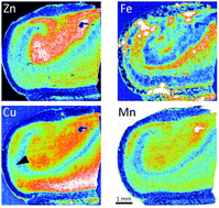
The loss of metal homeostasis has been implicated in the pathophysiology of mesial temporal lobe epilepsy associated with hippocampal sclerosis (mTLE-HS). Here we applied laser ablation inductively coupled plasma mass spectrometry imaging to establish the spatial distribution of Zn, Fe, Cu and Mn in coronal sections of hippocampi of four patients with drug-resistant mTLE-HS who underwent amygdalohippocampectomy. Detailed maps of the metal concentrations in the different morphological areas/layers were built and analyzed. The highest level of Zn (>20 μg g−1) was found in mossy fiber-rich regions – cornu ammonis field 4 (CA4), gyrus dentatus, and CA3. The distribution of Fe appears to reflect the routes of the main intrahippocampal blood vessels. The highest concentrations of Cu (>10 μg g−1) and Mn (>15 μg g−1) were observed in regions/layers with neuron somata – subiculum, CA4, gyrus dentatus, and stratum pyramidale (SPy) in CA1 and CA2. Alveus and other regions with axons and dendrites generally showed lower levels of Zn, Cu, and Mn. The Cu concentration was decreased in the areas of total neuronal loss in SPy in CA1 (9.73 ± 0.91 μg g−1), compared to the subiculum (13.32 ± 1.29 μg g−1; p = 0.043). The Cu and Mn concentrations correlated positively with neuron density in the SPy in CA1 (R = 0.629, p < 0.001; and R = 0.391, p = 0.004). These results provide a deeper insight into hippocampal metabolism of metals, and pave the road for identifying the components of the mechanism of epileptogenesis among Cu and Mn transporters and metalloproteins.
- This article is part of the themed collection: Zinc in the Biosciences
 Please wait while we load your content...