Murine cutaneous leishmaniasis investigated by MALDI mass spectrometry imaging†
Abstract
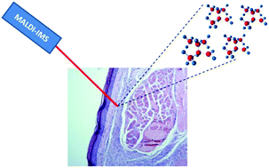
Imaging mass spectrometry (IMS) is recognized as a powerful tool to investigate the spatial distribution of untargeted or targeted molecules of a wide variety of samples including tissue sections. Leishmania is a protozoan parasite that causes different clinical manifestations in mammalian hosts. Leishmaniasis is a major public health risk in different continents and represents one of the most important neglected diseases. Cutaneous lesions from mice experimentally infected with Leishmania spp. were investigated by matrix-assisted laser desorption ionization MS using the SCiLS Lab software for statistical analysis. Being applied to cutaneous leishmaniasis (CL) for the first time, MALDI-IMS was used to search for peptides and low molecular weight proteins (2–10 kDa) as candidates for potential biomarkers. Footpad sections of Balb/c mice infected with (i) Leishmania amazonensis or (ii) Leishmania major were imaged. The comparison between healthy and infected skin highlighted a set of twelve possible biomarker proteins for L. amazonenis and four proteins for L. major. Further characterization of these proteins could reveal how these proteins act in pathology progression and confirm their values as biomarkers.


 Please wait while we load your content...