Label-free detection of aggregated platelets in blood by machine-learning-aided optofluidic time-stretch microscopy†‡

Abstract
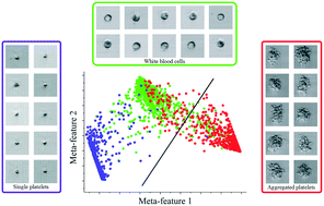
According to WHO, about 10 million new cases of thrombotic disorders are diagnosed worldwide every year. Thrombotic disorders, including atherothrombosis (the leading cause of death in the US and Europe), are induced by occlusion of blood vessels, due to the formation of blood clots in which aggregated platelets play an important role. The presence of aggregated platelets in blood may be related to atherothrombosis (especially acute myocardial infarction) and is, hence, useful as a potential biomarker for the disease. However, conventional high-throughput blood analysers fail to accurately identify aggregated platelets in blood. Here we present an in vitro on-chip assay for label-free, single-cell image-based detection of aggregated platelets in human blood. This assay builds on a combination of optofluidic time-stretch microscopy on a microfluidic chip operating at a high throughput of 10 000 blood cells per second with machine learning, enabling morphology-based identification and enumeration of aggregated platelets in a short period of time. By performing cell classification with machine learning, we differentiate aggregated platelets from single platelets and white blood cells with a high specificity and sensitivity of 96.6% for both. Our results indicate that the assay is potentially promising as predictive diagnosis and therapeutic monitoring of thrombotic disorders in clinical settings.
 Please wait while we load your content...

