Cytotoxic and sublethal effects of silver nanoparticles on tendon-derived stem cells – implications for tendon engineering
Abstract
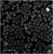
Tendon injuries occur commonly in sports and workplace. Tendon-derived stem cells (TDSCs) have great potential for tendon healing because they can differentiate into functional tenocytes. To grow TDSCs properly in vivo, a scaffold is needed. Silver nanoparticles (AgNPs) have been used in a range of biomedical applications for their anti-bacterial and -inflammatory effects. AgNPs are therefore expected to be a good scaffolding coating material for tendon engineering. Yet, their cytotoxicity in TDSCs remains unknown. Moreover, their sublethal effects were mysterious in TDSCs. In our study, decahedral AgNPs (43.5 nm in diameter) coated with polyvinylpyrrolidone (PVP) caused a decrease in TDSCs’ viability beginning at 37.5 μg ml−1 but showed non-cytotoxic effects at concentrations below 18.8 μg ml−1. Apoptosis was observed in the TDSCs when higher doses of AgNPs (75–150 μg ml−1) were used. Mechanistically, AgNPs induced reactive oxygen species (ROS) formation and mitochondrial membrane potential (MMP) depolarization, resulting in apoptosis. Interestingly, treating TDSCs with N-acetyl-L-cysteine (NAC) antioxidant significantly antagonized the ROS formation, MMP depolarization and apoptosis indicating that ROS accumulation was a prominent mediator in the AgNP-induced cytotoxicity. On the other hand, AgNPs inhibited the tendon markers’ mRNA expression (0–15 μg ml−1), proliferation and clonogenicity (0–15 μg ml−1) in TDSCs under non-cytotoxic concentrations. Taken together, we have reported here for the first time that the decahedral AgNPs are cytotoxic to rat TDSCs and their sublethal effects are also detrimental to stem cells’ proliferation and tenogenic differentiation. Therefore, AgNPs are not a good scaffolding coating material for tendon engineering.


 Please wait while we load your content...