Potential of silk fibroin/chondrocyte constructs of muga silkworm Antheraea assamensis for cartilage tissue engineering†
Abstract
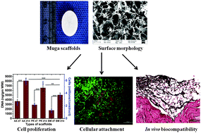
Articular cartilage damage represents one of the most perplexing clinical problems of musculoskeletal therapeutics due to its limited self-repair and regenerative capabilities. In this study, 3D porous silk fibroin scaffolds derived from non-mulberry muga silkworm Antheraea assamensis were fabricated and examined for their ability to support cartilage tissue engineering. Additionally, Bombyx mori and Philosamia ricini silk fibroin scaffolds were utilized for comparative studies. Herein, the fabricated scaffolds were thoroughly characterized and compared for cartilaginous tissue formation within the silk fibroin scaffolds seeded with primary porcine chondrocytes and cultured in vitro for 2 weeks. Surface morphology and structural conformation studies revealed the highly interconnected porous structure (pore size 80–150 μm) with enhanced stability within their structure. The fabricated scaffolds demonstrated improved mechanical properties and were followed-up with sequential experiments to reveal improved thermal and degradation properties. Silk fibroin scaffolds of A. assamensis and P. ricini supported better chondrocyte attachment and proliferation as indicated by metabolic activities and fluorescence microscopic studies. Biochemical analysis demonstrated significantly higher production of sulphated glycosaminoglycans (sGAGs) and type II collagen in A. assamensis silk fibroin scaffolds followed by P. ricini and B. mori scaffolds (p < 0.001). Furthermore, histochemistry and immunohistochemical studies indicated enhanced accumulation of sGAGs and expression of collagen II. Moreover, the scaffolds in a subcutaneous model of rat demonstrated in vivo biocompatibility after 8 weeks of implantation. Taken together, these results demonstrate the positive attributes from the non-mulberry silk fibroin scaffold of A. assamensis and suggest its suitability as a promising scaffold for chondrocyte based cartilage repair.


 Please wait while we load your content...