
3D scaffold induces efficient bone repair: in vivo studies of ultra-structural architecture at the interface
 e
and
e
and
Abstract
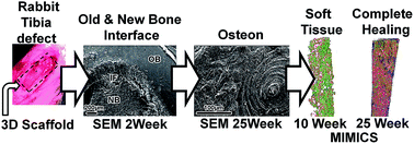
The repair of critical bone loss remains a challenge to orthopaedic surgeons. Various artificial scaffolds have been intensively evaluated to provide an alternative solution for the repair and regeneration of bone defects; however, the inconsistent clinical performances of available materials have prompted the development of reactive 3D scaffolds for bone tissue engineering. We have studied the ability of a functionally designed 3D scaffold to bridge critical size defects and induce new bone formation in a New Zealand white rabbit tibial model, and have evaluated its ultra-structural properties using a combination of techniques, such as solid-state nuclear magnetic resonance (ssNMR), scanning electron microscopy (SEM), energy dispersive X-ray (EDX) and micro-computed tomography (μ-CT) with MIMICS® (Materialise's Interactive Medical Image Control System). ssNMR showed the structural similarity of the synthetic biomaterial to naturally occurring human bone. SEM studies showed an increase in Ca/P ratio with time, the progressively uniform distribution of elements in healed bones, and increased new bone formation, finally resembling native (intact) bone. μ-CT and MIMICS® demonstrated the pattern and morphology of new bone formed, with a noticeable shift in the HU unit towards compact bone, from week 2 to 25. The results suggest that in the critical size bone defect, the scaffold enhanced the formation of new bone having biomaterial composition, ultra-structure and quality resembling that of native bone, thus suggesting significant improvement in guided bone regeneration. This research provides a promising new avenue for orthopaedic implant design that safely biodegrades while promoting new bone growth.
 Please wait while we load your content...

