The architecture of neutrophil extracellular traps investigated by atomic force microscopy†
Abstract
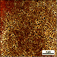
Neutrophils are immune cells that engage in a suicidal pathway leading to the release of partially decondensed chromatin, or neutrophil extracellular traps (NETs). NETs behave as a double edged sword; they can bind to pathogens thereby ensnaring them and limiting their spread during infection; however, they may bind to host circulating materials and trigger thrombotic events, and are associated with autoimmune disorders. Despite the fundamental role of NETs as part of an immune system response, there is currently a very poor understanding of how their nanoscale properties are reflected in their macroscopic impact. In this work, using a combination of fluorescence and atomic force microscopy, we show that NETs appear as a branching filament network that results in a substantially organized porous structure with openings with 0.03 ± 0.04 μm2 on average and thus in the size range of small pathogens. Topological profiles typically up to 3 ± 1 nm in height are compatible with a “beads on a string” model of nucleosome chromatin. Typical branch lengths of 153 ± 103 nm appearing as rigid rods and height profiles of naked DNA in NETs of 1.2 ± 0.5 nm are indicative of extensive DNA supercoiling throughout NETs. The presence of DNA duplexes could also be inferred from force spectroscopy and the occurrence of force plateaus that ranged from ∼65 pN to 300 pN. Proteolytic digestion of NETs resulted in widespread disassembly of the network structure and considerable loss of mechanical properties. Our results suggest that the underlying structure of NETs is considerably organized and that part of its protein content plays an important role in maintaining its mesh architecture. We anticipate that NETs may work as microscopic mechanical sieves with elastic properties that stem from their DNA–protein composition, which is able to segregate particles also as a result of their size. Such a behavior may explain their participation in capturing pathogens and their association with thrombosis.


 Please wait while we load your content...