MicroXRF tomographic visualization of zinc and iron in the zebrafish embryo at the onset of the hatching period†
Abstract
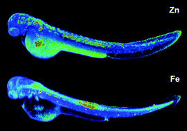
Transition metals such as zinc, copper, and iron play key roles in cellular proliferation, cell differentiation, growth, and development. Over the past decade, advances in synchrotron X-ray fluorescence instrumentation presented new opportunities for the three-dimensional mapping of trace metal distributions within intact specimens. Taking advantage of microXRF tomography, we visualized the 3D distribution of zinc and iron in a zebrafish embryo at the onset of the hatching period. The reconstructed volumetric data revealed distinct differences in the elemental distributions, with zinc predominantly localized to the yolk and yolk extension, and iron to various regions of the brain as well as the myotome extending along the dorsal side of the embryo. The data set complements an earlier tomographic study of an embryo at the pharyngula stage (24 hpf), thus offering new insights into the trace metal distribution at key stages of embryonic development.
- This article is part of the themed collections: RSC papers by GRC Metals in Medicine 2018 Speakers, Zinc in the Biosciences and Fifth International Symposium on Metallomics, Beijing, China
 Please wait while we load your content...