Nano-clustering of ligands on surrogate antigen presenting cells modulates T cell membrane adhesion and organization†
Abstract
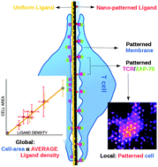
We investigate the adhesion and molecular organization of the plasma membrane of T lymphocytes interacting with a surrogate antigen presenting cell comprising glass supported ordered arrays of antibody (α-CD3) nano-dots dispersed in a non-adhesive matrix of polyethylene glycol (PEG). The local membrane adhesion and topography, as well as the distribution of the T cell receptors (TCRs) and the kinase ZAP-70, are influenced by dot-geometry, whereas the cell spreading area is determined by the overall average density of the ligands rather than specific characteristics of the dots. TCR clusters are recruited preferentially to the nano-dots and the TCR cluster size distribution has a weak dot-size dependence. On the patterns, the clusters are larger, more numerous, and more enriched in TCRs, as compared to the homogeneously distributed ligands at comparable concentrations. These observations support the idea that non-ligated TCRs residing in the non-adhered parts of the proximal membrane are able to diffuse and enrich the existing clusters at the ligand dots. However, long distance transport is impaired and cluster centralization in the form of a central supramolecular cluster (cSMAC) is not observed. Time-lapse imaging of early cell-surface contacts indicates that the ZAP-70 microclusters are directly recruited to the site of the antibody dots and this process is concomitant with membrane adhesion. These results together point to a complex interplay of adhesion, molecular organization and activation in response to spatially modulated stimulation.


 Please wait while we load your content...