Issue 59, 2016

Observation of intracellular interactions between DNA origami and lysosomes by the fluorescence localization method

Abstract

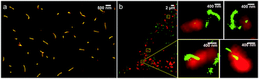
We obtained the fluorescence localization images of tube DNA origami nanostructures in NIH 3T3 cells for the first time. The fluorescence localization images of tube DNA origami nanostructures and TIRF images of lysosomes were combined and they revealed the detailed interactions between the two structures. Quantitative analysis illustrated that the tube origami can be captured as well as degraded by lysosomes with time.

Graphical abstract: Observation of intracellular interactions between DNA origami and lysosomes by the fluorescence localization method

Supplementary files

Article information

Article type
Communication
Submitted
18 Jan 2016
Accepted
19 Jun 2016
First published
20 Jun 2016

Chem. Commun., 2016,52, 9240-9242

Observation of intracellular interactions between DNA origami and lysosomes by the fluorescence localization method

M. Fu, L. Dai, Q. Jiang, Y. Tang, X. Zhang, B. Ding and J. Li, Chem. Commun., 2016, 52, 9240 DOI: 10.1039/C6CC00484A

To request permission to reproduce material from this article, please go to the Copyright Clearance Center request page.

If you are an author contributing to an RSC publication, you do not need to request permission provided correct acknowledgement is given.

If you are the author of this article, you do not need to request permission to reproduce figures and diagrams provided correct acknowledgement is given. If you want to reproduce the whole article in a third-party publication (excluding your thesis/dissertation for which permission is not required) please go to the Copyright Clearance Center request page.

Read more about how to correctly acknowledge RSC content.

Social activity

Spotlight

Advertisements